Teaching Cases in Advanced Breast Cancer: CT Imaging of Rare and Aggressive Presentations
Purpose or Learning Objective
The purpose of this work is to bring to attention to the importance of CT in evaluating the aggressiveness of breast cancer and to make women more aware of its development if neglected.
Breast cancer is the 2nd most common cancer worldwide and the number one cancer in women. Although it is a disease that can be easily prevented, a lot of women tend to neglect themselves and they present to the doctor in advanced stages. CT is very important in analyzing advanced stages of breast cancer.
Results and Findings
This series includes five female patients aged 51 to 78 years who presented to the Emergency Department for acute, non–breast-related symptoms. In all cases, contrast-enhanced CT performed for unrelated indications incidentally revealed previously undiagnosed malignancies, prompting further imaging and histopathological confirmation.
The detected pathologies are presented in order of rarity and educational value, with breast carcinomas grouped together.
Case 1 – Bilateral Breast Anaplastic Large Cell Lymphoma (ALCL) Without Implants
Bilateral breast anaplastic large cell lymphoma (ALCL) in a patient without breast implants represents the rarest and most exceptional case within this series of incidental CT detections. This presentation is remarkable due to the combination of three uncommon and clinically significant features: involvement of breast tissue by ALCL, a rare subtype of peripheral T‑cell lymphoma; the absence of any breast implants, excluding the more commonly described implant‑associated ALCL; and bilateral breast involvement at initial presentation, suggesting systemic disease with an unusual first localization.
ALCL is a subtype of non‑Hodgkin lymphoma characterized by pleomorphic large cells expressing CD30, and it has been most frequently associated with breast implants in recent literature. In contrast, breast ALCL without implants is exceedingly rare, with only isolated case reports and small case series described worldwide. Bilateral presentations in the absence of implants are even more exceptional, and this combination substantially broadens the differential diagnosis when encountered on imaging.
In this case, a contrast‑enhanced CT scan performed in the emergency setting for unrelated acute symptoms was the first modality to reveal bilateral enhancing soft‑tissue masses within the breast parenchyma. These masses were associated with enlarged regional lymph nodes, raising initial concern for either advanced epithelial malignancy or systemic lymphoproliferative disease. No peri‑implant fluid collections or signs of implant rupture were present, confirming the patient had no implants.
CT and Histology Highlights:
- Bilateral enhancing soft‑tissue masses in both breasts
- Associated regional lymphadenopathy without peri‑implant fluid
- No history of breast prostheses or prior breast interventions
- Biopsy: CD30‑positive anaplastic large cell lymphoma
- Exceptionally rare presentation, expanding the imaging differential for bilateral breast masses
Case 2 – Fibromyxoid Sarcoma
A female patient in her sixth decade presented to the Emergency Department with acute, non‑specific abdominal discomfort and flank pain, for which a contrast‑enhanced CT examination was ordered. While the initial clinical concern was unrelated to oncologic pathology, the CT images incidentally demonstrated a well‑defined soft‑tissue mass within the chest wall region, prompting further evaluation.
The mass exhibited heterogeneous enhancement following contrast administration, with internal areas of lower attenuation suggestive of myxoid stroma. There were no signs of adjacent bone destruction, neurovascular invasion, or distant metastases at the time of imaging. The radiologic appearance did not conform to common benign soft‑tissue lesions, raising suspicion for a rare malignant process. Given these atypical imaging features, the patient underwent image‑guided biopsy, and histopathological examination confirmed the diagnosis of fibromyxoid sarcoma.
CT and Histology Highlights:
- Well‑defined chest wall soft‑tissue mass with heterogeneous contrast enhancement
- Internal myxoid‑appearing areas of low attenuation
- Absence of overt bone, neurovascular, or distant metastatic involvement
- Biopsy confirmed fibromyxoid sarcoma, a rare soft‑tissue malignancy
- CT served as the first imaging modality to detect a clinically silent yet malignant lesion
Invasive Breast Carcinomas: 3 interesting cases
Histopathological analysis ultimately revealed two cases of invasive ductal carcinoma (IDC) and one case of invasive lobular carcinoma (ILC), representing the most common and second most common subtypes of invasive breast malignancy, respectively.
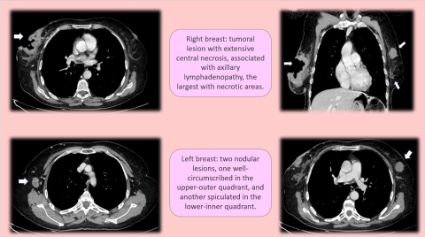
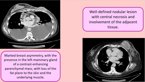
In the first of these cases, the patient’s contrast‑enhanced CT showed bilateral spiculated masses with heterogeneous enhancement within both breasts, accompanied by regional lymphadenopathy, peritoneal carcinomatosis, and multiple hepatic lesions. These findings indicated advanced systemic dissemination at first presentation, a scenario that underscores the power of CT to detect both primary malignancy and distant spread in a single examination. Subsequent targeted breast imaging and biopsy confirmed high‑grade invasive ductal carcinoma in both breasts.
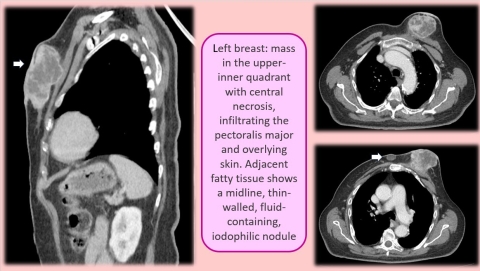
The second IDC case involved a unilateral enhancing breast mass with associated ipsilateral axillary lymphadenopathy, without evidence of distant metastatic disease. The radiologic appearance was consistent with invasive ductal carcinoma, and histopathology confirmed the diagnosis following directed biopsy.
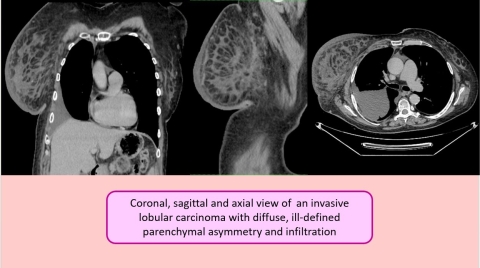
The patient with invasive lobular carcinoma demonstrated more subtle imaging features, with diffuse, ill‑defined parenchymal asymmetry and infiltration rather than a well‑circumscribed mass. This pattern is characteristic of the infiltrative “single‑file” growth of lobular carcinoma, which can be more difficult to recognize on imaging compared to ductal carcinoma. No distant metastases were identified in this case at the time of detection.
CT and Histology Highlights:
- Bilateral IDC: spiculated enhancing masses, regional lymphadenopathy, peritoneal carcinomatosis, hepatic metastases
- Unilateral IDC: enhancing mass with ipsilateral lymphadenopathy, no distant metastases
- ILC: diffuse breast parenchymal asymmetry and infiltration, no discrete mass, no metastases
- Biopsy confirmed all three invasive breast malignancies
- CT demonstrated capability to detect both primary tumors and advanced metastatic disease
Conclusion
CT imaging has an important role in assessing advanced breast cancer due to its accurate evaluation of local invasion and metastatic disease.